CellPaint TSP膜染色液
 |
货号 |
22700 |
存储条件 |
在零下15度以下保存, 避免光照 |
| 规格 |
500 Tests |
价格 |
2604 |
| Ex (nm) |
496 |
Em (nm) |
633 |
| 分子量 |
1016.27 |
溶剂 |
DMSO |
| 产品详细介绍 |
简要概述
产品基本信息
货号:22700
产品名称:CellPaint TSP膜染色液
规格:500 Tests
储存条件:-15℃避光防潮
保质期:12个月
产品物理化学光谱特性
分子量:1016.27
外观:固体
溶剂:DMSO
激发波长(nm):N/A
发射波长(nm):N/A
适用仪器
| 荧光显微镜 |
|
| 激发: |
Cy3/TRITC滤波片 |
| 发射: |
Cy3/TRITC滤波片 |
| 推荐孔板: |
黑色透明 |
产品介绍
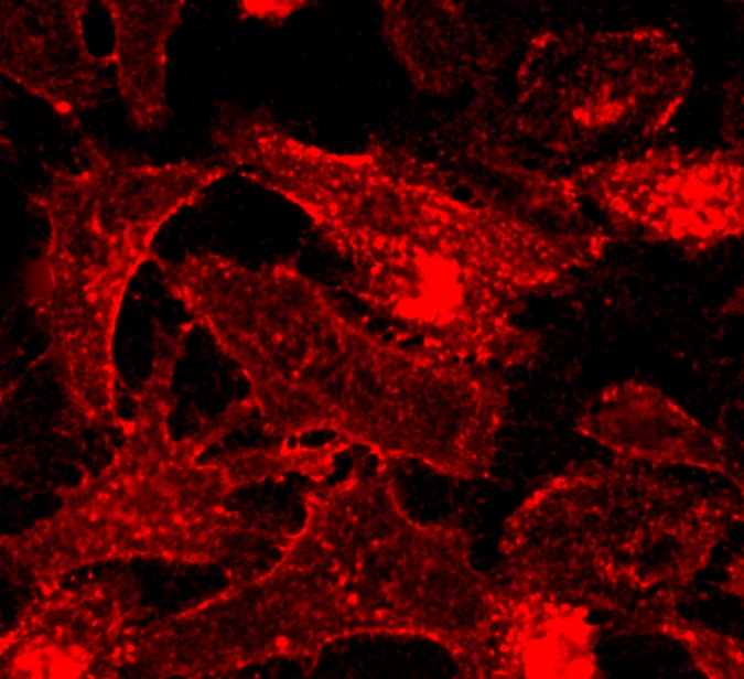
TSP是一种基于苯乙烯基吡啶的荧光膜探针,适用于活体细胞和组织的质膜成像。TSP探针是一个分子转子,在粘性介质中荧光急剧增强,它的荧光对微环境也很敏感,能够对高信噪比的质膜进行成像。TSP光稳定性高、细胞毒性低和生物相容性良好。它也可用于双光子成像。金畔生物是AAT Bioquest的中国代理商,为您提供最优质的CellPaint TSP膜染色液。
点击查看光谱
参考文献
Membrane trafficking and exocytosis are upregulated in port wine stain blood vessels
Authors: Yin, R., Rice, S. J., Wang, J., Gao, L., Tsai, J., Anvari, R. T., Zhou, F., Liu, X., Wang, G., Tang, Y., Mihm, M. C., Jr., Belani, C. P., Chen, D. B., Nelson, J. S., Tan, W.
Journal: Histol Histopathol (2019): 479-490
Cu(2+)-Directed Liposome Membrane Fusion, Positive-Stain Electron Microscopy, and Oxidation
Authors: Liu, Y., Liu, J.
Journal: Langmuir (2018): 7545-7553
Extraction of DNA from Sperm Cells in Mixed Stain by Nylon Membrane Bushing Separation Technique
Authors: Ma, J., Tong, Q., Gao, L. B., Zhu, C., Jiang, Z. Q.
Journal: Fa Yi Xue Za Zhi (2018): 417-419
Gram’s Stain Does Not Cross the Bacterial Cytoplasmic Membrane
Authors: Wilhelm, M. J., Sheffield, J. B., Sharifian Gh, M., Wu, Y., Spahr, C., Gonella, G., Xu, B., Dai, H. L.
Journal: ACS Chem Biol (2015): 1711-7
The use of SMALPs as a novel membrane protein scaffold for structure study by negative stain electron microscopy
Authors: Postis, V., Rawson, S., Mitchell, J. K., Lee, S. C., Parslow, R. A., Dafforn, T. R., Baldwin, S. A., Muench, S. P.
Journal: Biochim Biophys Acta (2015): 496-501
Overview of electron crystallography of membrane proteins: crystallization and screening strategies using negative stain electron microscopy
Authors: Nannenga, B. L., Iadanza, M. G., Vollmar, B. S., Gonen, T.
Journal: Curr Protoc Protein Sci (2013): Unit17 15
Negative-stain electron microscopy of inside-out FtsZ rings reconstituted on artificial membrane tubules show ribbons of protofilaments
Authors: Milam, S. L., Osawa, M., Erickson, H. P.
Journal: Biophys J (2012): 59-68
Ultrastructure and retinal imaging of internal limiting membrane: a clinicopathologic correlation of trypan blue stain in macular hole surgery
Authors: Mackenzie, S. E., G and orfer, A., Rohleder, M., Schumann, R., Schlottmann, P. G., Bunce, C., Xing, W., Gregor, Z., Charteris, D. G.
Journal: Retina (2010): 655-61
A nitrobenzofuran-conjugated phosphatidylcholine (C12-NBD-PC) as a stain for membrane lamellae for both microscopic imaging and spectrofluorimetry
Authors: Hope-Roberts, M., Wainwright, M., Horobin, R. W.
Journal: Biotech Histochem (2008): 25-8
Indocyanine green-assisted internal limiting membrane peeling for macular holes to stain or not to stain?
Authors: Da Mata, A. P., Riemann, C. D., Nehemy, M. B., Foster, R. E., Petersen, M. R., Burk, S. E.
Journal: Retina (2005): 395-404
说明书
CellPaint TSP膜染色液.pdf


