上海金畔生物科技有限公司提供具砂板存储球闪式层析柱,欣维尔,C394625C,可以访问官网了解更多产品信息。
具砂板存储球闪式层析柱,欣维尔,C394625C
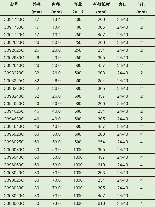
产品简介:本品系国外普遍使用的低压闪式层析柱,每个层析柱标配一个四氟节门塞。
技术参数:
| 材质 | 玻璃 |
| 规格(mm) | 254 |
| 标准口 | 24/40 |
| 砂芯 | |
| 活塞 |

上海金畔生物科技有限公司提供具砂板存储球闪式层析柱,欣维尔,C394625C,可以访问官网了解更多产品信息。
具砂板存储球闪式层析柱,欣维尔,C394625C
产品简介:本品系国外普遍使用的低压闪式层析柱,每个层析柱标配一个四氟节门塞。
技术参数:

| 材质 | 玻璃 |
| 规格(mm) | 254 |
| 标准口 | 24/40 |
| 砂芯 | |
| 活塞 |
上海金畔生物科技有限公司代理AAT Bioquest荧光染料全线产品,欢迎访问AAT Bioquest荧光染料官网了解更多信息。
 |
货号 | 50028 | 存储条件 | 在零下15度以下保存 |
| 规格 | 1 mg | 价格 | 6564 | |
| Ex (nm) | Em (nm) | |||
| 分子量 | 溶剂 | |||
| 产品详细介绍 | ||||
简要概述
产品基本信息
货号:50028
产品名称:磺胺噻唑-HRP缀合物
规格:1mg
储存条件:-15℃避光防潮
保质期:12个月
产品介绍
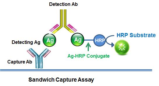
磺胺噻唑-HRP缀合物用于开发ELISA测定法,用于定量磺胺噻唑。 磺胺噻唑-HRP缀合物也可用于定量磺胺噻唑,磺胺甲恶唑,磺胺甲恶唑和磺胺异恶唑。 磺胺噻唑-HRP共轭物在每个HRP上具有最少数目的磺胺噻唑分子,以使其测定灵敏度最大化。金畔生物是AAT Bioquest的中国代理商,为您提供最优质的磺胺嘧啶-HRP缀合物。
参考文献
A high-throughput, precipitating colorimetric sandwich ELISA microarray for shiga toxins
Authors: Gehring A, He X, Fratamico P, Lee J, Bagi L, Brewster J, Paoli G, He Y, Xie Y, Skinner C, Barnett C, Harris D.
Journal: Toxins (Basel) (2014): 1855
Development of antigen capture ELISA for the quantification of EIAV p26 protein
Authors: Hu Z, Chang H, Ge M, Lin Y, Wang X, Guo W.
Journal: Appl Microbiol Biotechnol (2014): 9073
Development of atom transfer radical polymer-modified gold nanoparticle-based enzyme-linked immunosorbent assay (ELISA)
Authors: Chen F, Hou S, Li Q, Fan H, Fan R, Xu Z, Zhala G, Mai X, Chen X, Liu Y.
Journal: Anal Chem (2014): 10021
Development of monoclonal antibody-based sandwich ELISA for detection of dextran
Authors: Wang SY, Li Z, Wang XJ, Lv S, Yang Y, Zeng LQ, Luo FH, Yan JH, Liang DF.
Journal: Monoclon Antib Immunodiagn Immunother (2014): 334
Effect of different combinations of antibodies and enzyme labels on ELISA of progesterone
Authors: Kumari GL, P and ey PK, Nathsharma SS, Sharma SK, Kochhar G.
Journal: J Immunoassay Immunochem (2014): 157
Long-term dry storage of an enzyme-based reagent system for ELISA in point-of-care devices
Authors: Ramach and ran S, Fu E, Lutz B, Yager P.
Journal: Analyst (2014): 1456
Ultrasensitive ELISA using enzyme-loaded nanospherical brushes as labels
Authors: Qu Z, Xu H, Xu P, Chen K, Mu R, Fu J, Gu H.
Journal: Anal Chem (2014): 9367
3-(10′-Phenothiazinyl)propionic acid is a potent primary enhancer of peroxidase-induced chemiluminescence and its application in sensitive ELISA of methylglyoxal-modified low density lipoprotein
Authors: Sakharov IY, Demiyanova AS, Gribas AV, Uskova NA, Efremov EE, Vdovenko MM.
Journal: Talanta (2013): 414
Development of an ELISA for the quantification of the C-terminal decapeptide prothymosin alpha(100-109) in sera of mice infected with bacteria
Authors: Samara P, Kalbacher H, Ioannou K, Radu DL, Livaniou E, Promponas VJ, Voelter W, Tsitsilonis O.
Journal: J Immunol Methods (2013): 54
ELISA for determination of total coagulation factor XII concentration in human plasma
Authors: Madsen DE, Sidelmann JJ, Overgaard K, Koch C, Gram JB.
Journal: J Immunol Methods (2013): 32
上海金畔生物科技有限公司可以定制不同序列多肽,可以访问官网了解更多产品信息。
| 名称 | pTH (1-84) (human) |
| 编码 | [68893-82-3] |
| 别名 | pTH (1-84) (human) |
| 纯度 | 80%,90%,95%,98%,99% |
| 重量 | 1mg,5mg,10mg,50mg,100mg,1g |
| 序列(单字母缩写) | SVSEIQLMHNLGKHLNSMERVEWLRKKLQDVHNFVALGAPLAPRDAGSQRPRKKEDNVLVESHEKSLGEADKADVNVLTKAKSQ |
| 序列(三字母缩写) | Ser-Val-Ser-Glu-Ile-Gln-Leu-Met-His-Asn-Leu-Gly-Lys-His-Leu-Asn-Ser-Met-Glu-Arg-Val-Glu-Trp-Leu-Arg-Lys-Lys-Leu-Gln-Asp-Val-His-Asn-Phe-Val-Ala-Leu-Gly-Ala-Pro-Leu-Ala-Pro-Arg-Asp-Ala-Gly-Ser-Gln-Arg-Pro-Arg-Lys-Lys-Glu-Asp-Asn-Val-Leu-Val-Glu-Ser-His-Glu-Lys-Ser-Leu-Gly-Glu-Ala-Asp-Lys-Ala-Asp-Val-Asn-Val-Leu-Thr-Lys-Ala-Lys-Ser-Gln |
| 基本描述 | |
| 溶解度 | |
| 分子量 | 0 |
| 化学式 | |
| 存储条件 | Store at -20°C. Keep tightly closed. Store in a cool dry place. |
| 注释 | |
| Documents | ![pTH (1-84) (human) 编码 [68893-82-3]](http://www.saliva.com.cn/wp-content/uploads/2022/10/20221005_633d29bf9b574.png) |
| Figures | ![pTH (1-84) (human) 编码 [68893-82-3]](http://www.saliva.com.cn/wp-content/uploads/2022/10/20221005_633d29bff1cfd.jpg) |
| Reference | |
| C端 | |
| N端 | |
| 化学桥 |
上海金畔生物科技有限公司提供具砂板存储球闪式层析柱,欣维尔,C394620C,可以访问官网了解更多产品信息。
具砂板存储球闪式层析柱,欣维尔,C394620C
产品简介:本品系国外普遍使用的低压闪式层析柱,每个层析柱标配一个四氟节门塞。
技术参数:

| 材质 | 玻璃 |
| 规格(mm) | 203 |
| 标准口 | 24/40 |
| 砂芯 | |
| 活塞 |
上海金畔生物科技有限公司代理AAT Bioquest荧光染料全线产品,欢迎访问AAT Bioquest荧光染料官网了解更多信息。
| 货号 | 985 | 存储条件 | 在零下15度以下保存, 避免光照 | |
| 规格 | 1 mg | 价格 | 2604 | |
| Ex (nm) | 789 | Em (nm) | 814 | |
| 分子量 | 799.05 | 溶剂 | DMSO | |
| 产品详细介绍 | ||||
简要概述
产品基本信息
货号:985
产品名称:吲哚菁绿ICG叠氮化物
规格:1mg
储存条件:-15℃避光防潮
保质期:12个月
产品物理化学光谱特性
分子量:799.05
外观:固体
溶剂:DMSO
激发波长(nm):780
发射波长(nm):800
产品介绍
吲哚菁绿(ICG)是一种三碳菁型染料,具有NIR吸收特性(峰值吸收约780 nm),大发射波长约为800 nm。FDA批准了非侵入性近红外(NIR)荧光成像染料ICG进行眼科血管造影,以确定心输出量以及肝血流量和功能。由于红外频率穿透视网膜层,因此与荧光素血管造影相比,ICG血管造影可以成像更深的循环模式。 ICG与血浆蛋白紧密结合,并局限于血管系统。 ICG的半衰期为150到180秒,只能从肝脏转移至胆汁而从循环系统中移除。近的一项研究表明,ICG可在注射后20分钟内靶向动脉粥样硬化,并为体内检测动脉粥样硬化兔中富含脂质,发炎,冠状动脉粥样斑块提供了足够的信号增强。离体荧光反射成像显示,与注射盐水的带有粥样斑块的兔子相比,注射了ICG的带有粥样斑块的兔子的斑块靶标与背景比率高。它还可用于其他医学诊断和癌症患者中,用于实体瘤的检测,淋巴结的定位以及在重建手术期间的血管造影,视网膜和脉络膜血管的可视化以及光动力疗法。在癌症诊断和治疗中,ICG既可以用作显像染料又可以用作热疗剂。在可见光范围内几乎没有吸收,这是由于低自发荧光,组织吸收和在近红外波长(700-900 nm)下的散射引起的。这种ICG叠氮化物可用于通过众所周知的点击化学化学选择性标记炔烃标记的生物分子(如蛋白质,脂质,核酸,糖)。金畔生物是AAT Bioquest的中国代理商,为您提供优质的吲哚菁绿ICG叠氮化物。
点击查看光谱
参考文献
Assessment of Lexiscan for Blood Brain Barrier disruption to facilitate Fluorescence brain imaging
Authors: Pak, Rebecca W and Le, Hanh and Valentine, Heather and Thorek, Daniel and Rahmim, Arman and Wong, Dean and Kang, Jin U
Journal: (2017): ATu3B–2
Bioengineering of injectable encapsulated aggregates of pluripotent stem cells for therapy of myocardial infarction
Authors: Zhao, Shuting and Xu, Zhaobin and Wang, Hai and Reese, Benjamin E and Gushchina, Liubov V and Jiang, Meng and Agarwal, Pranay and Xu, Jiangsheng and Zhang, Mingjun and Shen, Rulong and others
Journal: Nature Communications (2016): 13306
Deep Photoacoustic/Luminescence/Magnetic Resonance Multimodal Imaging in Living Subjects Using High-Efficiency Upconversion Nanocomposites
Authors: Liu, Yu and Kang, Ning and Lv, Jing and Zhou, Zijian and Zhao, Qingliang and Ma, Lingceng and Chen, Zhong and Ren, Lei and Nie, Liming
Journal: Advanced Materials (2016)
Single-Layer MoS2 Nanosheets with Amplified Photoacoustic Effect for Highly Sensitive Photoacoustic Imaging of Orthotopic Brain Tumors
Authors: Chen, Jingqin and Liu, Chengbo and Hu, Dehong and Wang, Feng and Wu, Haiwei and Gong, Xiaojing and Liu, Xin and Song, Liang and Sheng, Zonghai and Zheng, Hairong
Journal: Advanced Functional Materials (2016)
A simple and effective technique for identification of intersegmental planes by infrared thoracoscopy after transbronchial injection of indocyanine green
Authors: Sekine Y, Ko E, Oishi H, Miwa M.
Journal: J Thorac Cardiovasc Surg. (2012)
Adverse Reaction in Patients with Drug Allergy History After Simultaneous Intravenous Fundus Fluorescein Angiography and Indocyanine Green Angiography
Authors: Su Z, Ye P, Teng Y, Zhang L, Shu X.
Journal: J Ocul Pharmacol Ther. (2012)
An innovative method for detecting surgical errors using indocyanine green angiography during carotid endarterectomy: a preliminary investigation
Authors: Lee CH, Jung YS, Yang HJ, Son YJ, Lee SH.
Journal: Acta Neurochir (Wien) (2012): 67
Anatomic response of occult choroidal neovascularization to intravitreal ranibizumab: a study by indocyanine green angiography
Authors: Querques G, Tran TH, Forte R, Querques L, B and ello F, Souied EH.
Journal: Graefes Arch Clin Exp Ophthalmol (2012): 479
Applicability of radiocolloids, blue dyes and fluorescent indocyanine green to sentinel node biopsy in melanoma
Authors: Uhara H, Yamazaki N, Takata M, Inoue Y, Sakakibara A, Nakamura Y, Suehiro K, Yamamoto A, Kamo R, Mochida K, Takenaka H, Yamashita T, Takenouchi T, Yoshikawa S, Takahashi A, Uehara J, Kawai M, Iwata H, Kadono T, Kai Y, Watanabe S, Murata S, Ikeda T, Fukamizu H, Tanaka T, Hatta N, Saida T.
Journal: J Dermatol (2012): 336
Application of indocyanine green videoangiography in surgery for spinal vascular malformations
Authors: Misra BK, Pur and are HR.
Journal: J Clin Neurosci. (2012)
上海金畔生物科技有限公司可以定制不同序列多肽,可以访问官网了解更多产品信息。
| 名称 | pTH (1-84) (rat) |
| 编码 | |
| 别名 | pTH (1-84) (rat) |
| 纯度 | 80%,90%,95%,98%,99% |
| 重量 | 1mg,5mg,10mg,50mg,100mg,1g |
| 序列(单字母缩写) | AVSEIQLMHNLGKHLASVERMQWLRKKLQDVHNFVSLGVQMAAREGSYQRPTKKEENVLVDGNSKSLGEGDKADVDVLVKAKSQ |
| 序列(三字母缩写) | Ala-Val-Ser-Glu-Ile-Gln-Leu-Met-His-Asn-Leu-Gly-Lys-His-Leu-Ala-Ser-Val-Glu-Arg-Met-Gln-Trp-Leu-Arg-Lys-Lys-Leu-Gln-Asp-Val-His-Asn-Phe-Val-Ser-Leu-Gly-Val-Gln-Met-Ala-Ala-Arg-Glu-Gly-Ser-Tyr-Gln-Arg-Pro-Thr-Lys-Lys-Glu-Glu-Asn-Val-Leu-Val-Asp-Gly-Asn-Ser-Lys-Ser-Leu-Gly-Glu-Gly-Asp-Lys-Ala-Asp-Val-Asp-Val-Leu-Val-Lys-Ala-Lys-Ser-Gln |
| 基本描述 | |
| 溶解度 | |
| 分子量 | 0 |
| 化学式 | |
| 存储条件 | Store at -20°C. Keep tightly closed. Store in a cool dry place. |
| 注释 | |
| Documents |  |
| Figures |  |
| Reference | |
| C端 | |
| N端 | |
| 化学桥 |
上海金畔生物科技有限公司提供具砂板存储球闪式层析柱,欣维尔,C393240C,可以访问官网了解更多产品信息。
具砂板存储球闪式层析柱,欣维尔,C393240C
产品简介:本品系国外普遍使用的低压闪式层析柱,每个层析柱标配一个四氟节门塞。
技术参数:

| 材质 | 玻璃 |
| 规格(mm) | 457 |
| 标准口 | 24/40 |
| 砂芯 | |
| 活塞 |
上海金畔生物科技有限公司代理AAT Bioquest荧光染料全线产品,欢迎访问AAT Bioquest荧光染料官网了解更多信息。
 |
货号 | 20089 | 存储条件 | 在零下15度以下保存, 避免光照 |
| 规格 | 100 tests | 价格 | 2604 | |
| Ex (nm) | 410 | Em (nm) | 455 | |
| 分子量 | ~36000 | 溶剂 | Water | |
| 产品详细介绍 | ||||
简要概述
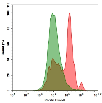
Annexin V,PacBlue缀合物是美国AAT Bioquest研发的用于检测细胞功能的探针,膜联蛋白是钙依赖性磷脂结合蛋白家族。 它们富含真核生物,属于参与信号转导的普遍存在的细胞质蛋白家族。 膜联蛋白V的优先结合配偶体是磷脂酰丝氨酸(PS),其通常保持在细胞膜的内叶(细胞质侧)。 在细胞凋亡中,PS被转移到质膜的外部小叶。 磷脂酰丝氨酸在细胞表面上的出现是细胞凋亡的初始/中间阶段的通用指示,并且可以在观察到形态变化之前检测到。 我们的膜联蛋白V结合物是一组监测细胞功能的工具,旨在通过测量磷脂酰丝氨酸的易位来监测细胞凋亡。
膜联蛋白V结合物在Ca2+存在下与凋亡细胞表面上的PS结合,它也可以通过坏死细胞膜或死细胞并与细胞内部的PS结合。 因此,我们建议将细胞不可渗透的核染色与膜联蛋白V结合物结合使用,以区分死细胞和凋亡细胞。金畔生物是AAT Bioquest的中国代理商,为您提供最优质的Annexin V,PacBlue缀合物。
产品说明书
分析方案
概述
用测试化合物(200μL/样品)制备细胞
添加Annexin V结合物测定溶液
室温孵育30-60分钟
用流式细胞仪或荧光显微镜分析
1.用膜联蛋白V缀合物制备和孵育细胞:
1.1制备膜联蛋白V结合测定缓冲液:10mM HEPES,140mM NaCl和2.5mM CaCl2,pH 7.4。
1.2用测试化合物处理细胞一段所需的时间(用星形孢菌素处理的Jurkat细胞4-6小时)以诱导细胞凋亡。
1.3离心细胞,得到1-5×10⁵个细胞/管。
1.4将细胞重悬于200μL膜联蛋白V结合测定缓冲液中(来自步骤1.1)。
1.5在细胞中加入2μL膜联蛋白V结合物。
可选:在细胞内加入死细胞染色剂如碘化丙锭,用于坏死细胞。
1.6在室温下孵育30至60分钟,避光。
1.7在用流式细胞仪或荧光显微镜分析细胞之前,加入300μL膜联蛋白V结合测定缓冲液(来自步骤1.1)以增加体积(参见下面的步骤1.8)。
1.8使用流式细胞仪或荧光显微镜监测荧光强度(参见下面的步骤2或3)。
2.使用流式细胞仪分析:
通过使用具有适当过滤器的流式细胞仪来量化膜联蛋白V缀合物。
注意:粘附细胞上的膜联蛋白V结合流式细胞术分析未经常检测,因为在细胞分离或收获期间可能发生特定的膜损伤。 然而,Casiola-Rosen等人先前报道了利用膜联蛋白V对贴壁细胞类型进行流式细胞术的方法。 和van Engelend等人(见参考文献1和2)。
3.使用荧光显微镜分析:
3.1移取步骤1.6的细胞悬液,用膜联蛋白V结合测定缓冲液(来自步骤1.1)冲洗1-2次,然后用膜联蛋白V结合测定缓冲液(来自步骤1.1)重悬细胞。 将细胞添加到盖有玻璃盖玻片的载玻片上。
注意:对于粘附细胞,建议直接在盖玻片上生长细胞。 与膜联蛋白V缀合物孵育(步骤1.6)后,用膜联蛋白V结合测定缓冲液(来自步骤1.1)冲洗1-2次,并将膜联蛋白V结合测定缓冲液(来自步骤1.1)加回到盖玻片中。 在玻璃载玻片上翻转盖玻片并观察细胞。 在与膜联蛋白V缀合物孵育后,细胞也可以在2%甲醛中固定,并在显微镜下观察。
3.2在荧光显微镜下用适当的滤光片分析膜联蛋白V缀合物的凋亡细胞
参考文献
White light-induced cell apoptosis by a conjugated polyelectrolyte through singlet oxygen generation
Authors: Jiamei Liang, Pan Wu, Chunyan Tan, Yuyang Jiang
Journal: RSC advances (2018): 9218–9222
Resources-Application Notes Phenotypic and Epigenetic Mechanism of Action Determinations of Histone Methylase and Demethylase Inhibitors using Digital Widefield Microscopy
Authors: Brad Larson, Peter Banks
Journal: Experimental Biology (2017)
Targeted Magnetic Intra-Lysosomal Hyperthermia produces lysosomal reactive oxygen species and causes Caspase-1 dependent cell death
Authors: Pascal Clerc, Pauline Jeanjean, Nicolas Halalli, Michel Gougeon, Bernard Pipy, Julian Carrey, Daniel Fourmy, Véronique Gigoux
Journal: Journal of Controlled Release (2017)
Detecting Apoptosis, Autophagy, and Necrosis
Authors: Jack Coleman, Rui Liu, Kathy Wang, Arun Kumar
Journal: (2016): 77–92
相关产品
| 产品名称 | 货号 |
| Annexin V-mFluor Violet 540标记 | Cat#20082 |
| Annexin V-mFluor Violet 450标记 | Cat#20080 |
| Annexin V-Cy7标记 | Cat#20068 |
上海金畔生物科技有限公司可以定制不同序列多肽,可以访问官网了解更多产品信息。
| 名称 | pTH (18-48) (human) |
| 编码 | [153238-99-4] |
| 别名 | pTH (18-48) (human) |
| 纯度 | 80%,90%,95%,98%,99% |
| 重量 | 1mg,5mg,10mg,50mg,100mg,1g |
| 序列(单字母缩写) | MERVEWLRKKLQDVHNFVALGAPLAPRDAGS-OH |
| 序列(三字母缩写) | Met-Glu-Arg-Val-Glu-Trp-Leu-Arg-Lys-Lys-Leu-Gln-Asp-Val-His-Asn-Phe-Val-Ala-Leu-Gly-Ala-Pro-Leu-Ala-Pro-Arg-Asp-Ala-Gly-Ser |
| 基本描述 | |
| 溶解度 | |
| 分子量 | 0 |
| 化学式 | |
| 存储条件 | Store at -20°C. Keep tightly closed. Store in a cool dry place. |
| 注释 | |
| Documents | ![pTH (18-48) (human) 编码 [153238-99-4]](http://www.saliva.com.cn/wp-content/uploads/2022/10/20221005_633d29afa2f92.png) |
| Figures | ![pTH (18-48) (human) 编码 [153238-99-4]](http://www.saliva.com.cn/wp-content/uploads/2022/10/20221005_633d29b000165.jpg) |
| Reference | |
| C端 | |
| N端 | |
| 化学桥 |
上海金畔生物科技有限公司提供具砂板存储球闪式层析柱,欣维尔,C393230C,可以访问官网了解更多产品信息。
具砂板存储球闪式层析柱,欣维尔,C393230C
产品简介:本品系国外普遍使用的低压闪式层析柱,每个层析柱标配一个四氟节门塞。
技术参数:

| 材质 | 玻璃 |
| 规格(mm) | 305 |
| 标准口 | 24/40 |
| 砂芯 | |
| 活塞 |
上海金畔生物科技有限公司代理AAT Bioquest荧光染料全线产品,欢迎访问AAT Bioquest荧光染料官网了解更多信息。
 |
货号 | 22280 | 存储条件 | 在零下15度以下保存, 避免光照 |
| 规格 | 500 Tests | 价格 | 2604 | |
| Ex (nm) | 415 | Em (nm) | 499 | |
| 分子量 | 557.01 | 溶剂 | DMSO | |
| 产品详细介绍 | ||||
简要概述
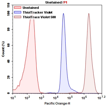
ThiolTrace Violet 500*谷胱甘肽检测试剂*是美国AAT Bioquest生产的荧光染料,GSH的亚细胞检测和定位对于了解氧化还原状态的调节,药物的作用以及排毒的机制非常重要。ThiolTrace Violet 500是一种比通常使用的mBBr,mBCl或Thiotracker Violet更亮,更坚固的细胞内硫醇探针,可用于监测细胞内GSH。由于还原型谷胱甘肽代表细胞中细胞内大多数游离巯基,因此ThiolTrace 紫罗兰500可用于估算还原型谷胱甘肽的细胞水平。它比mBCL和其他细胞内常见的硫醇检测探针(例如Thiotracker紫罗兰色)至少亮10倍,并且可以通过紫外线或405 nm的大斯托克斯位移进行激发。它可以用醛固定,并通过Triton®X-100(0.5%)渗透。它可以用于包括细胞毒性研究在内的多重分析中。ThiolTrace Violet 500提供了一种简单,灵敏且可重现的工具来检测生物样品中降低的GSH含量。ThiolTrace Violet 500与硫醇反应,在普通的405 nm紫激光下激发出520-530 nm的强荧光。与ThiolTracker紫罗兰(Thermo Fisher Scientific)相比,ThiolTrace紫罗兰500在含有生长因子的细胞培养基中的强度高10-100倍。ThiolTrace Violet 500与多种稀释剂兼容,包括含血清的细胞培养基。ThiolTrace Violet 500可用于流式细胞仪,HCS成像和落射荧光显微镜。
点击查看光谱
适用仪器
| 流式细胞仪 | |
| 激发: | 405nm激光 |
| 发射: | 525/50nm滤波片 |
| 通道: | Pacific Orange通道 |
| 荧光显微镜 | |
| 激发: | 405nm |
| 发射: | 525nm |
| 推荐孔板: | 黑色透明 |
| 滤波片: | TRITC滤波片组 |
产品说明书
样品实验方案
简要概述
1.用5×10 5到1×10 6细胞/ mL 的密度制备含测试化合物的细胞
2.准备ThiolTrace Violet 500工作溶液并将其添加到细胞中
3.在37 o C 孵育20至30分钟
4.在Ex / Em = 405/525 nm处读取荧光强度-Pacific Orange
溶液配制
1.储备溶液配制
所有未使用的储备溶液应分为一次性使用的等分试样,并在制备后储存在-20°C下。 避免重复冻融循环。
ThiolTrace 紫罗兰色500储备液(500X):
在小瓶ThiolTrace Violet 500中加入200 µL DMSO(未提供),并充分混合。 注意:分装并在-20℃下存储未使用的ThiolTrace Violet 500储备液。 避免重复冷冻/融化循环。
2.工作溶液配制
ThiolTrace Violet 500工作溶液(1X):
将1 µL ThiolTraceTM紫罗兰色500储备溶液加入您选择的0.5 mL缓冲液中,并充分混合。 注意:可以在含有血清的细胞培养基中制备ThiolTrace Violet 500工作溶液。
点击查看细胞样品制备
操作步骤
1.用测试化合物处理细胞所需的时间。注意:对于贴壁细胞,用0.5 mM EDTA轻轻提起细胞以保持细胞完整,并在用ThiolTrace 紫罗兰色工作溶液孵育之前,用含血清的培养基洗涤细胞一次。注意:适当的孵育时间取决于所用的单个细胞类型和细胞浓度。优化每个实验的孵育时间。
2.将细胞以1000 rpm的速度离心4分钟,然后用1 mL所选缓冲液(可选)洗涤细胞。
3.将细胞重悬于0.5 mL ThiolTraceTM Violet 500工作溶液中,并在37oC培养箱中孵育20至30分钟。注意:对于荧光显微镜,每孔添加200 µL ThiolTraceTM Violet 500工作溶液。
4.以1000 rpm离心细胞4分钟,然后在1 mL所选缓冲液(可选)中洗涤细胞。
5.重悬于缓冲液中,使用太平洋橙滤光片组(Ex / Em = 405/525 nm),用流式细胞仪监测荧光强度。
参考文献
A highly sensitive two-photon fluorescent probe for glutathione with near-infrared emission at 719nm and intracellular glutathione imaging
Authors: C. Huang
Journal: Spectrochim Acta A Mol Biomol Spectrosc (2019): 68-76
Glutathione-driven Cu(i)-O2 chemistry: a new light-up fluorescent assay for intracellular glutathione
Authors: P. F. Gao
Journal: Analyst (2018): 2486-2490
Nitrogen-doped carbon quantum dots as a fluorescent probe to detect copper ions, glutathione, and intracellular pH
Authors: S. Liao
Journal: Anal Bioanal Chem (2018): 7701-7710
Graphene Quantum Dot-MnO2 Nanosheet Based Optical Sensing Platform: A Sensitive Fluorescence “Turn Off-On” Nanosensor for Glutathione Detection and Intracellular Imaging
Authors: X. Yan
Journal: ACS Appl Mater Interfaces (2016): 21990-6
A sensitive turn-on fluorescent probe for intracellular imaging of glutathione using single-layer MnO2 nanosheet-quenched fluorescent carbon quantum dots
Authors: D. He
Journal: Chem Commun (Camb) (2015): 14764-7
A fluorescent probe for intracellular cysteine overcoming the interference by glutathione
Authors: M. Tian
Journal: Org Biomol Chem (2014): 6128-33
A mitochondria-targetable fluorescent probe for dual-channel NO imaging assisted by intracellular cysteine and glutathione
Authors: Y. Q. Sun
Journal: J Am Chem Soc (2014): 12520-3
Designing an intracellular fluorescent probe for glutathione: two modulation sites for selective signal transduction
Authors: M. Isik
Journal: Org Lett (2014): 3260-3
Turn-on fluorescence sensor for intracellular imaging of glutathione using g-C(3)N(4) nanosheet-MnO(2) sandwich nanocomposite
Authors: X. L. Zhang
Journal: Anal Chem (2014): 3426-34
Detection of intracellular glutathione using ThiolTracker violet stain and fluorescence microscopy
Authors: B. S. Mandavilli
Journal: Curr Protoc Cytom (2010): Unit 9 35
上海金畔生物科技有限公司可以定制不同序列多肽,可以访问官网了解更多产品信息。
| 名称 | pTH (2-38) (human) |
| 编码 | |
| 别名 | pTH (2-38) (human) |
| 纯度 | 80%,90%,95%,98%,99% |
| 重量 | 1mg,5mg,10mg,50mg,100mg,1g |
| 序列(单字母缩写) | VSEIQLMHNLGKHLNSMERVEWLRKKLQDVHNFVALG-OH |
| 序列(三字母缩写) | Val-Ser-Glu-Ile-Gln-Leu-Met-His-Asn-Leu-Gly-Lys-His-Leu-Asn-Ser-Met-Glu-Arg-Val-Glu-Trp-Leu-Arg-Lys-Lys-Leu-Gln-Asp-Val-His-Asn-Phe-Val-Ala-Leu-Gly |
| 基本描述 | |
| 溶解度 | |
| 分子量 | 0 |
| 化学式 | |
| 存储条件 | Store at -20°C. Keep tightly closed. Store in a cool dry place. |
| 注释 | |
| Documents |  |
| Figures |  |
| Reference | |
| C端 | |
| N端 | |
| 化学桥 |
上海金畔生物科技有限公司提供具砂板存储球闪式层析柱,欣维尔,C393225C,可以访问官网了解更多产品信息。
具砂板存储球闪式层析柱,欣维尔,C393225C
产品简介:本品系国外普遍使用的低压闪式层析柱,每个层析柱标配一个四氟节门塞。
技术参数:

| 材质 | 玻璃 |
| 规格(mm) | 254 |
| 标准口 | 24/40 |
| 砂芯 | |
| 活塞 |
上海金畔生物科技有限公司代理AAT Bioquest荧光染料全线产品,欢迎访问AAT Bioquest荧光染料官网了解更多信息。
| 货号 | 1821 | 存储条件 | 在零下15度以下保存, 避免光照 |
| 规格 | 1 mg | 价格 | 2604 |
| Ex (nm) | 553 | Em (nm) | 568 |
| 分子量 | ~1250 | 溶剂 | DMSO |
| 产品详细介绍 | |||
简要概述
产品基本信息
货号:1821
产品名称:AF555 NHS酯类
规格:1mg
储存条件:保存在冰箱-15℃干燥
保质期:12个月
产品物理化学光谱特性
分子量:~1250
外观:固体
溶剂:DMSO
激发波长(nm):555
发射波长(nm):572
产品介绍
AF555 NHS酯(琥珀酰亚胺酯)与AlexaFluor®555NHS酯相同(AlexaFluor®是ThermoFisher的商标)。它是一种鲜红色的荧光染料。AF555染料是水溶性的并且pH从pH4到pH10不敏感。AF555的NHS酯(或琥珀酰亚胺酯)是最方便的胺反应形式,用于将该染料与蛋白质或抗体结合。
点击查看光谱
点击查看实验方案
参考文献
A combined solvatochromic shift and TDDFT study probing solute-solvent interactions of blue fluorescent Alexa Fluor 350 dye: Evaluation of ground and excited state dipole moments
Authors: M. K. Patil
Journal: Spectrochim Acta A Mol Biomol Spectrosc (2019): 142-152
Photobleaching Comparison of R-Phycoerythrin-Streptavidin and Streptavidin-Alexa Fluor 568 in a Breast Cancer Cell Line
Authors: S. N. Ostad
Journal: Monoclon Antib Immunodiagn Immunother (2019): 25-29
Comparison between photostability of Alexa Fluor 448 and Alexa Fluor 647 with conventional dyes FITC and APC by flow cytometry
Authors: S. Rai
Journal: Int J Lab Hematol (2018): e52-e54
Development of new hCaM-Alexa Fluor((R)) biosensors for a wide range of ligands
Authors: I. Velazquez-Lopez
Journal: Anal Biochem (2017): 13-22
[Neuroanatomical basis of clinical joint application of “Jinggu” (BL 64, a source-acupoint) and “Dazhong” (KI 4, a Luo-acupoint) in the rat: a double-labeling study of cholera toxin subunit B conjugated with Alexa Fluor 488 and 594]
Authors: J. J. Cui
Journal: Zhen Ci Yan Jiu (2011): 262-7
[Neuroanatomical characteristics of acupoint “Chengshan” (BL 57) in the rat: a cholera toxin subunit B conjugated with Alexa Fluor 488 method study]
Authors: X. L. Zhu
Journal: Zhen Ci Yan Jiu (2010): 433-7
相关产品
| 产品名称 | 货号 |
| iFluor 555琥珀酰亚胺酯 | Cat#1028 |
上海金畔生物科技有限公司提供具砂板存储球闪式层析柱,欣维尔,C393220C,可以访问官网了解更多产品信息。
具砂板存储球闪式层析柱,欣维尔,C393220C
产品简介:本品系国外普遍使用的低压闪式层析柱,每个层析柱标配一个四氟节门塞。
技术参数:

| 材质 | 玻璃 |
| 规格(mm) | 203 |
| 标准口 | 24/40 |
| 砂芯 | |
| 活塞 |
上海金畔生物科技有限公司可以定制不同序列多肽,可以访问官网了解更多产品信息。
| 名称 | pTH (28-48) (human) |
| 编码 | [83286-22-0] |
| 别名 | pTH (28-48) (human) |
| 纯度 | 80%,90%,95%,98%,99% |
| 重量 | 1mg,5mg,10mg,50mg,100mg,1g |
| 序列(单字母缩写) | LQDVHNFVALGAPLAPRDAGS-OH |
| 序列(三字母缩写) | Leu-Gln-Asp-Val-His-Asn-Phe-Val-Ala-Leu-Gly-Ala-Pro-Leu-Ala-Pro-Arg-Asp-Ala-Gly-Ser |
| 基本描述 | |
| 溶解度 | |
| 分子量 | 0 |
| 化学式 | |
| 存储条件 | Store at -20°C. Keep tightly closed. Store in a cool dry place. |
| 注释 | |
| Documents | ![pTH (28-48) (human) 编码 [83286-22-0]](http://www.saliva.com.cn/wp-content/uploads/2022/10/20221005_633d299f8b76e.png) |
| Figures | ![pTH (28-48) (human) 编码 [83286-22-0]](http://www.saliva.com.cn/wp-content/uploads/2022/10/20221005_633d299fe04de.jpg) |
| Reference | |
| C端 | |
| N端 | |
| 化学桥 |
上海金畔生物科技有限公司代理AAT Bioquest荧光染料全线产品,欢迎访问AAT Bioquest荧光染料官网了解更多信息。
 |
货号 | 21800 | 存储条件 | 在零下15度以下保存, 避免光照 |
| 规格 | 1 mg | 价格 | 3264 | |
| Ex (nm) | 494 | Em (nm) | 521 | |
| 分子量 | 822.72 | 溶剂 | DMSO | |
| 产品详细介绍 | ||||
简要概述
CytoTrace Ultra Green新一代荧光示踪探针,非常适合跟踪细胞的运动或位置。该染料可以很好地保留在细胞中,并且追踪活细胞数代。与在相同条件下的CMFDA相比,CytoTrace Ultra Green明显更亮,光稳定性更高并且更耐用。在组织固定后,这种染料产生的信号将非常稳定,使其成为与其他类型的分析方法(如细胞毒性等)结合用于多色应用的理想选择。 CytoTrace Ultra Green的激发光谱和发射光谱与FITC相同,并且与常见的红色荧光染料(例如Texas Red,Cy5,Cy7,iFluor 647和750,AlexaFluor®647和750)完全分离。 CytoTrace Ultra Green可轻松用于各种生物应用的活细胞跟踪,并与流式细胞仪和荧光显微镜兼容。金畔生物是AAT Bioquest的中国代理商,为您提供最优质的CytoTrace Ultra Green。
产品说明书
实验方案
简要概述
溶液配制
储备溶液配制
CytoTrace Ultra Green原液(2-10 mM):添加适量的DMSO充分混合以制成CytoTrace Ultra Green原液(2-10 mM)。注意:储备溶液应立即使用;所有剩余的溶液应等分,并在<-20 o C下冷冻。避免重复冻融循环,并避光。
工作溶液配制
CytoTrace Ultra Green工作溶液:通过用Hanks和20 mM Hepes缓冲液(HHBS)或自备的pH=7的缓冲液稀释DMSO储备液,制备0.5至5 µM的染料工作液,并离心。注意:在某些细胞类型,可能需要较低的浓度才能染色细胞。我们建议在进行实验之前针对每种细胞类型进行测试以获得最佳浓度使实验更顺利。
操作步骤
图示
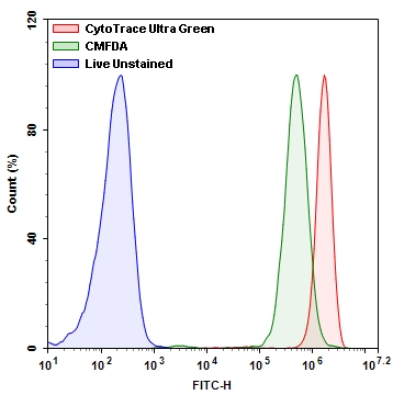
图1. Jurkat细胞中CytoTrace Ultra Green与CMFDA的荧光强度比较。在37 ℃,5%CO2培养箱中,将Jurkat细胞用CytoTrace Ultra Green和CMFDA染料分别加载30分钟。使用带有FITC通道的ACEA NovoCyte 3000流式细胞仪测量荧光强度。
参考文献
Fluorescence-Based Transport Assays Revisited in a Human Renal Proximal Tubule Cell Line
Authors: Caetano-Pinto P, Janssen MJ, Gijzen L, Verscheijden L, Wilmer MJ, Masereeuw R.
Journal: Mol Pharm (2016): 933
The variable chemotherapeutic response of Malabaricone-A in leukemic and solid tumor cell lines depends on the degree of redox imbalance
Authors: Manna A, De Sarkar S, De S, Bauri AK, Chattopadhyay S, Chatterjee M.
Journal: Phytomedicine (2015): 713
Cell membrane tracker based on restriction of intramolecular rotation
Authors: Zhang C, Jin S, Yang K, Xue X, Li Z, Jiang Y, Chen WQ, Dai L, Zou G, Liang XJ.
Journal: ACS Appl Mater Interfaces (2014): 8971
A multiple model probability hypothesis density tracker for time-lapse cell microscopy sequences
Authors: Rezatofighi SH, Gould S, Vo BN, Mele K, Hughes WE, Hartley R.
Journal: Inf Process Med Imaging (2013): 110
Evaluation of stability and sensitivity of cell fluorescent labels when used for cell migration
Authors: Beem E, Segal MS.
Journal: J Fluoresc (2013): 975
TLM-Tracker: software for cell segmentation, tracking and lineage analysis in time-lapse microscopy movies
Authors: Klein J, Leupold S, Biegler I, Biedendieck R, Munch R, Jahn D.
Journal: Bioinformatics (2012): 2276
Horizontal DNA transfer from donor to host cells as an alternative mechanism of epithelial chimerism after allogeneic hematopoietic cell transplantation
Authors: Waterhouse M, Themeli M, Bertz H, Zoumbos N, Finke J, Spyridonidis A.
Journal: Biol Blood Marrow Transplant (2011): 319
The exocytosis of fluorescent nanodiamond and its use as a long-term cell tracker
Authors: Fang CY, Vaijayanthimala V, Cheng CA, Yeh SH, Chang CF, Li CL, Chang HC.
Journal: Small (2011): 3363
The interplay between Leishmania promastigotes and human Natural Killer cells in vitro leads to direct lysis of Leishmania by NK cells and modulation of NK cell activity by Leishmania promastigotes
Authors: Lieke T, Nylen S, Eidsmo L, Schmetz C, Berg L, Akuffo H.
Journal: Parasitology (2011): 1898
Cell electrofusion visualized with fluorescence microscopy
Authors: Trontelj K, Usaj M, Miklavcic D.
Journal: J Vis Exp. (2010)
相关产品
| 货号 # | 产品名称 | 规格 | 分子量 | Ex/Em (nm) | 溶剂 |
| 22014 | CytoTrace Orange CMTMR | 10×50 mg | 554.04 | 541/565 | DMSO |
| 22015 | CytoTrace Red CMPTX | 10×50 mg | 686.25 | 577/602 | DMSO |
| 22016 | CytoTrace Red CFDA | 1 mg | 652.43 | 560/574 | DMSO |
| 22017 | CytoTrace Green CMFDA | 1 mg | 464.86 | 494/521 | DMSO |
| 21800 | CytoTrace Ultra Green | 1 mg | 822.72 | 494/521 | DMSO |
| 22020 | FDA (Fluorescein diacetate) | 1 g | 416.83 | 494/521 | DMSO |
上海金畔生物科技有限公司提供具砂板存储球闪式层析柱,欣维尔,C392640C,可以访问官网了解更多产品信息。
具砂板存储球闪式层析柱,欣维尔,C392640C
产品简介:本品系国外普遍使用的低压闪式层析柱,每个层析柱标配一个四氟节门塞。
技术参数:

| 材质 | 玻璃 |
| 规格(mm) | 457 |
| 标准口 | 24/40 |
| 砂芯 | |
| 活塞 |
上海金畔生物科技有限公司可以定制不同序列多肽,可以访问官网了解更多产品信息。
| 名称 | pTH (29-32) (human) |
| 编码 | [157879-49-8] |
| 别名 | pTH (29-32) (human) |
| 纯度 | 80%,90%,95%,98%,99% |
| 重量 | 1mg,5mg,10mg,50mg,100mg,1g |
| 序列(单字母缩写) | QDVH-OH |
| 序列(三字母缩写) | Gln-Asp-Val-His |
| 基本描述 | |
| 溶解度 | |
| 分子量 | 0 |
| 化学式 | |
| 存储条件 | Store at -20°C. Keep tightly closed. Store in a cool dry place. |
| 注释 | |
| Documents | ![pTH (29-32) (human) 编码 [157879-49-8]](http://www.saliva.com.cn/wp-content/uploads/2022/10/20221005_633d2997a6a44.png) |
| Figures | ![pTH (29-32) (human) 编码 [157879-49-8]](http://www.saliva.com.cn/wp-content/uploads/2022/10/20221005_633d299808400.jpg) |
| Reference | |
| C端 | |
| N端 | |
| 化学桥 |
上海金畔生物科技有限公司代理AAT Bioquest荧光染料全线产品,欢迎访问AAT Bioquest荧光染料官网了解更多信息。
 |
货号 | 20018 | 存储条件 | 在零下15度以下保存, 避免光照 |
| 规格 | 100 tests | 价格 | 2604 | |
| Ex (nm) | Em (nm) | |||
| 分子量 | ~36000 | 溶剂 | Water | |
| 产品详细介绍 | ||||
简要概述
Annexin V-生物素缀合物是美国AAT Bioquest生产的膜联蛋白,膜联蛋白是在钙存在下与磷脂膜结合的蛋白质家族。膜联蛋白V是研究细胞凋亡的有用工具。它被用作探针以检测在细胞表面上表达磷脂酰丝氨酸的细胞,这是细胞凋亡中发现的特征以及其他形式的细胞死亡。有多种参数可用于监测细胞活力。膜联蛋白V-染料缀合物广泛用于通过测量磷脂酰丝氨酸(PS)的易位来检测细胞凋亡。在细胞凋亡中,PS被转移到质膜的外部小叶。磷脂酰丝氨酸在细胞表面上的出现是细胞凋亡的初始/中间阶段的通用指示,并且可以在观察到形态变化之前检测到。Annexin V缀合物被广泛用于通过测量磷脂酰丝氨酸(PS)的转运来监测细胞凋亡。金畔生物是AAT Bioquest 的中国代理商,为您提供最优质的Annexin V-生物素缀合物。
产品说明书
操作步骤
1.用膜联蛋白V缀合物制备和孵育细胞:
1.1 制备膜联蛋白V缀合测定缓冲液:10mM HEPES,140mM NaCl和2.5mM CaCl2,pH 7.4。
1.2 用测试化合物处理细胞一段时间(用星形孢菌素处理的Jurkat细胞4-6小时)以诱导细胞凋亡。
1.3 离心细胞,得到1-5×10 5个细胞/管。
1.4 将细胞重悬于200μLAnnexin V缀合测定缓冲液中(来自步骤1.1)。
1.5 在细胞中加入2μL膜联蛋白V缀合物。
可选:在细胞内加入死细胞染色剂如碘化丙锭,用于死细胞。
1.6 在室温下进行30至60分钟,避光。
1.7 在用流式细胞仪或荧光显微镜分析细胞之前,加入300μLAnnexin V缀合测定缓冲液(来自步骤1.1)以增加体积(参见下面的步骤1.8)。
1.8 使用流式细胞仪或荧光显微镜检测荧光强度(参见下面的步骤2或3)。
2.使用流式细胞仪分析:
通过使用具有适当滤波器的流式细胞仪来量化膜联蛋白V缀合物。
注意:对贴壁细胞的膜联蛋白V结合流式细胞术分析未经过常规测试,因为在细胞分离过程中可能会发生特定膜损伤。
3.使用荧光显微镜分析:
3.1 吸取步骤1.6中的细胞悬液,用膜联蛋白V结合测定缓冲液(步骤1.1)冲洗1-2次,然后用膜联蛋白V结合测定缓冲液(步骤1.1)重悬细胞。 将细胞添加到载玻片上并盖上盖玻片。
注意:对于贴壁细胞,建议直接在盖玻片上培养细胞。 与膜联蛋白V结合物孵育后(步骤1.6),用膜联蛋白V结合测定缓冲液(来自步骤1.1)冲洗1-2次,然后将膜联蛋白V结合测定缓冲液(来自步骤1.1)加回到盖玻片上。 将盖玻片倒在载玻片上并使细胞可视化。 与膜联蛋白V缀合物一起孵育后,也可以将细胞固定在2%甲醛中,并在显微镜下观察。
3.2 在荧光显微镜下用适当的滤光片分析膜联蛋白V缀合物的凋亡细胞。
参考文献
Response of Mammalian cells to non-thermal intense narrowband pulsed electric fields
Authors: Sunao Katsuki, Yulan Li, Daiki Miyakawa, Ryo Yamada, Nobuaki Onishi, Soowon Lim
Journal: (2017): 1358–1361
相关产品
| 产品名称 | 货号 |
| Annexin V, TRITC标记 | Cat#20031 |